
 檢測認證人脈交流通訊錄
美國CIRS 134200牙科口腔模體詳細描述
美國CIRS 134200牙科口腔模體用于口腔內放射學評估,測試和評估(PIRATE)的CIRS幻影是頜骨診斷放射學的參考標準。幻影旨在幫助技術和臨床人員選擇,監測,培訓和驗證口腔內放射成像程序和其他常見牙科手術常見的掃描參數。
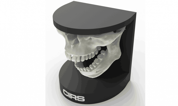
PIRATE幻影由上頜骨,下頜骨和保持裝置組成。幻影可以安裝到包含三腳架的球頭配件中,以實現定位的靈活性。上頜骨和下頜骨鉸接以允許牙科技術人員放置帶有保持器的眩光膜或其他口腔內裝置,并對幻影進行成像。
幻影由專有的組織等效材料構成,通過牙科X光和其他牙科成像程序的線性衰減模擬1%內的參考組織。
134200型近似于大小和結構上的平均男性人類下巴。幻影具有詳細的3D擬人解剖學,包括骨,竇,上頜骨,下頜骨,下頜神經和牙齒。骨骼包含皮層和小梁分離。牙齒包括不同的牙質,牙釉質,根管和牙冠。一個牙齒包括骨折,另一個牙齒包括腔。
美國CIRS 134200牙科口腔模體特點:
詳細的解剖特征
組織當量為50keV至25MeV
功能下顎
三腳架有六自由度
軟側手提箱
48個月的保修期
美國CIRS 134200牙科口腔模體應用:
口腔內X線放射治療儀
了解如何正確定位最佳圖像
用于種植體計劃和頜面重建的測試重建技術和算法
在實施新設備和技術時對人員進行培訓和評估
驗證圖像質量的一致性
The CIRS Phantom for Intraoral Radiography Assessment, Testing & Evaluation (PIRATE) is a standard of reference for diagnostic radiology of the jaw. The phantom is designed to assist technical and clinical staff in the selection, monitoring, training and verification of scanning parameters common to intraoral radiological imaging procedures and other common dental procedures.
The PIRATE phantom consists of maxilla, mandible and holding device. The phantom can be mounted to included tripod with ballhead fitting for flexibility in positioning. The maxilla and mandible are hinged to allow a dental technician to place a bitewing film or other intraoral devices with a holder and image the phantom.
The phantom is constructed of proprietary tissue-equivalent materials that mimic reference tissues within 1% by linear attenuation for the dental x-ray and other dental imaging procedures.
The Model 134200 approximates the average male human jaw in both size and structure. The phantom features detailed 3D anthropomorphic anatomy including bone, sinus, maxilla, mandible, mandibular nerve and teeth. The bones contain both cortical and trabecular separation. The teeth include distinct dentine, enamel, root canal and crown. One tooth includes a fracture and a second includes a cavity.
-
Detailed anatomical features
-
Tissue Equivalent from 50 keV to 25 MeV
-
Functioning Mandible
- Tripod with six degrees-of-freedom
-
Soft-sided carry case
-
48-month warranty
-
Commission intraoral X-ray devices
-
Learn how to properly position for optimal images
-
Test reconstruction techniques and algorithms for implant planning and maxillofacial reconstruction
-
Train and evaluate personnel during implementation of new equipment and techniques
-
Validate consistency of image quality
-